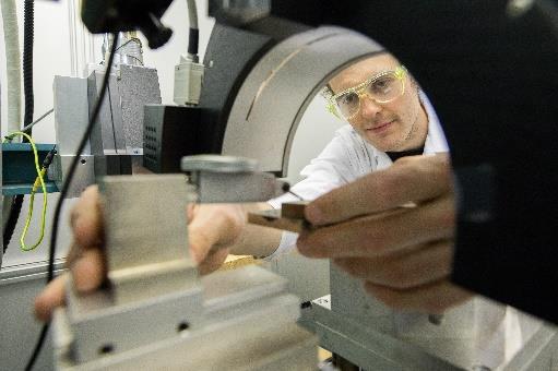
X-ray diffraction is a nondestructive measurement method based on the diffraction of X-rays in crystal lattices. Factors such as the crystal structures of the substances present, the crystallinity, the crystallite size, the crystallographic texture, the residual stresses and the purity influence the obtained measurement result. As manifold as the influencing factors are the measuring methods for their determination. At CEST there are four different measuring devices, optimized for special applications, in particular for the investigation of thin layers (GIXD).
Applications:
- Phase analysis – qualitative and quantitative, of both solid and powder samples
- Surface Analysis
- Residual stress measurements
- Texture measurements
- Particle size measurements
- Crystallinity measurements of polymers
Specifications:
- Euler cradle for determinations of the crystallographic texture as well as for residual stress measurements
- ω-goniometer for powder intake according to Bragg-Brentano and for grazing incidence measurements
- Image Plate Guinier camera in Seeman-Bohlin geometry for measuring thin layers and for “quasi” in situ observation of electrode processes
- Huber θ-θ Multipurpose Diffractometer with Fast Image Plate Detection (STOE) and PSD System (STOE)
Additional Equipment:
- Accessory unit for reflection measurements
Applications:
- In situ measurements (during electrochemical treatment)
- Materials Analysis
- Quality control
- Process Development
- Corrosion Analysis
- Texture measurements
- Residual stress measurements
- Qualitative phase analyzes
- Quantitative phase analyzes
Sample requirements:
- Solid or powder
- Solid samples should be as flat as possible and not particularly rough
- Size up to 30 x 30 cm
- Height up to 20 cm


